
Home > Research Achievements > Structural mechanism of the regulation of ADP release from molecular motor kinesin and implication for the kinesin motility
Structural mechanism of the regulation of ADP release from molecular motor kinesin and implication for the kinesin motility
(Nature Struct. Mol. Biol. 15: 1067-75, 2008)
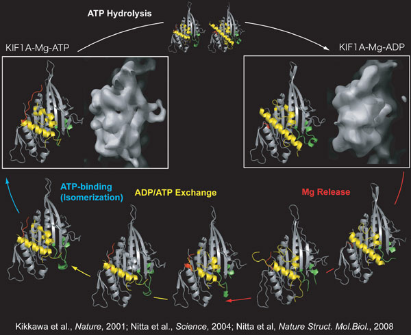
Molecular motor kinesin plays a critical role in the intracellular transport of various organelles, protein complexes and mRNAs.To elucidate the molecular mechanism of the kinesin motility, we have solved the crystal and electron microscopic structures of monomeric kinesin KIF1A in the various intermediate states during the ATP hydrolysis cycle.Here we solved the series of crystal structures during the release of ADP so that we have successfully solved all intermediate structures during the ATP hydrolysis cycle (Fig. 1).
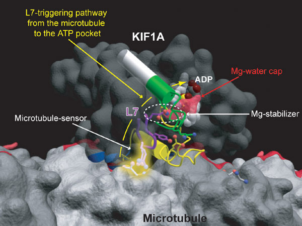
ADP release is the rate-limiting slow step of KIF1A, and the microtubule, the rail of KIF1A, accelerates this step more than 10,000 times.The molecular mechanism of this slow release is as follows.ADP in the ATP pocket is covered and stabilized by the dense hydrogen-bond network of magnesium ion and its very stable coordinated waters (Fig. 2).This “Mg-water cap” is connected to the root of the rigid β-hairpin structure L7 through the linkage namely “Mg-stabilizer”, which further stabilize ADP.This tight trap of ADP results in the very slow release of ADP.Meanwhile, the tip of L7 (microtubule-sensor) is exposed on the microtubule-binding surface.Microtubule recognizes this “microtubule-sensor” and pulls it toward the microtubule by the electrostatic interaction.The “Mg-stabilizer” breaks first, the “Mg-water cap” is then released, and finally the ADP is released.By this L7-triggering strategy, KIF1A can effectively use the energy of ATP only when it is on the rail and saves energy.Program member
Nobutaka Hirokawa (Department of Cell Biology & Anatomy, Graduate School of Medicine)
Copyright (C) 2007-2009 All rights reserved tokyo-u.ac.jp